A large bruise does not tell you whether an eye injury is dangerous, and a small eyelid cut can damage the tear-drainage system. You will learn which signs require emergency treatment, which injuries need same-day ophthalmic or oculoplastic review, and when scheduled observation is reasonable after examination.
Key takeaways
- Go to the emergency department for reduced vision, a displaced eye or a new RAPD.
- Ask an eyelid injury specialist to assess cuts involving the lid margin, tear duct or muscle.
- Observe orbital fractures only after vision, eye movement and globe position are documented.
- Arrange follow-up if swelling, double vision, pain or vision changes worsen after discharge.
Which facial-trauma signs mean you should go to the emergency department?
Do not decide that it is safe to wait from the size of a bruise or cut. When facial trauma needs oculoplastic care in Chennai depends on the injured structure and eye findings: reduced vision, a new relative afferent pupillary defect, or a displaced eye demands urgent assessment.
- Go to the emergency department for an irregular or peaked pupil, suspected globe penetration, severe eye pain, a firm or displaced eye, proptosis, restricted eye movement, new double vision, or rising eye pressure.
- Go immediately for uncontrolled bleeding, facial numbness, restricted jaw opening, or clear fluid leaking from the nose.
- Treat rapidly worsening pain or swelling, tense eyelids, increasing protrusion, vision loss, ophthalmoplegia, nausea or vomiting, faintness, bradycardia, or a tense orbit as possible orbital compartment syndrome. Waiting for bruising to settle can cost vision.
- If an open globe is possible, place a rigid eye shield over the eye, avoid pressure or manipulation, do not explore or suture the wound, and seek emergency ophthalmic care.
| Finding | Appropriate action | Why waiting is unsafe |
|---|---|---|
| Emergency eye signs | Emergency department now | Vision, globe integrity, or orbital pressure may be threatened |
| Complex but stable injury | Same-day ophthalmology or oculoplastic review | Eyelid, tear duct, fracture, or nerve damage needs examination |
| Clean superficial injury with normal documented findings | Planned observation with return precautions | Delayed swelling, infection, or double vision can change the plan |
What happens during the first assessment, and which level of care is appropriate?
The first assessment decides whether you need an emergency department, same-day ophthalmology or oculoplastic review, or documented outpatient observation. Clinicians record visual acuity, pupil responses, eye movements, globe position, facial sensation, and intraocular pressure only when measuring it is safe.
Slit-lamp examination checks the cornea and anterior eye; eyelid inspection covers the skin, lid margins, canthi, tarsus, levator region, and tear ducts.
| Level of care | What it means | When it applies |
|---|---|---|
| Emergency department | Immediate ophthalmic and trauma assessment | Suspected open globe, reduced vision, new relative afferent pupillary defect, irregular or peaked pupil, severe pain, proptosis, restricted movement, rapidly worsening swelling, tense eyelids, or a firm orbit |
| Same-day specialist review | Prompt ophthalmology or oculoplastic examination | New double vision, facial numbness, a deep eyelid wound, medial canthal injury, suspected canalicular damage, tissue loss, or a possible orbital fracture |
| Planned observation | Recorded findings with named follow-up and return precautions | Clean superficial cut or stable fracture after normal eye examination, irrigation, and appropriate closure |
CT of the facial bones and orbits shows fracture lines, trapped muscle, foreign bodies, sinus involvement, and displaced soft tissue, but it cannot replace the eye examination. The wound still needs irrigation, a retained-foreign-body check, tetanus review, and antibiotics when contamination, a bite, an open fracture, or lacrimal-system repair makes infection prevention relevant.
That is the basis for deciding on oculoplastic treatment after facial injury in Chennai—not the bruise’s size.
Which eyelid cuts need an eyelid injury specialist?
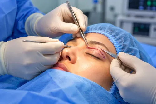
Normal vision does not clear an eyelid wound: the globe may be intact while the lid’s alignment, drainage, support, or lifting muscle is damaged. A superficial cut can be routine; anatomy matters more than length.
| Injury | Why specialist assessment matters | Usual consequence if missed |
|---|---|---|
| Superficial skin cut | Spares the lid margin, medial canthus, canaliculus, tarsus, levator, and globe | Irrigation and closure after eye examination may suffice |
| Full-thickness lid-margin laceration | The margin must be aligned precisely | Notching, lash misdirection, or corneal exposure |
| Medial canthal injury or canalicular tear | The tear-drainage channel may be divided | Persistent epiphora unless the canaliculus is identified and stented |
| Tarsal-plate or levator injury | Lid support or elevation is disrupted | Malposition, incomplete closure, or drooping |
| Tissue loss or contamination | Reconstruction and staged wound control may be needed | Infection, poor healing, or exposure |
A medial eyelid wound is treated as a possible canalicular injury until assessed. Closing only the skin can seal the surface while leaving the drainage channel disconnected; repair commonly requires locating the canaliculus and placing a stent.
Deep, contaminated, or anatomically complex wounds should reach an eyelid injury specialist in Chennai for urgent repair or planned reconstruction based on tissue damage and contamination.
A clean cut outside these structures still needs irrigation, a retained-foreign-body check, tetanus review, and antibiotics when bites, open fractures, heavy contamination, or lacrimal repair make them relevant.
A peaked pupil, prolapsed tissue, or penetrating-object history suggests open-globe injury: place a rigid shield, avoid pressure, and seek emergency ophthalmic care rather than explore or suture it.
When does an orbital fracture need surgery rather than observation?
A CT finding alone does not mandate orbital-fracture surgery. A small, stable orbital floor fracture can be observed when vision is intact, there is no muscle entrapment, no persistent functionally significant double vision, no troublesome globe displacement, and no visual impairment.
Use these three pathways:
- Immediate repair: Seek emergency care for vision-threatening compression, an open contaminated injury, or muscle entrapment causing nausea, vomiting, bradycardia, faintness, or severe restriction of eye movement. A child with a trapdoor fracture and marked movement restriction needs urgent assessment; waiting for bruising to fade can risk permanent muscle injury.
- Repair after swelling settles: Adults can discuss delayed repair once edema improves if disabling diplopia remains in primary or reading gaze, clinically important enophthalmos or hypoglobus develops, or the orbital defect is large. Roughly 2 mm of enophthalmos is a commonly cited reference point, not an automatic surgical rule.
- Observation with reassessment: Arrange documented follow-up rather than relying on photographs or appearance. Return promptly for worsening pain or swelling, reduced vision, new double vision, increasing protrusion, fever, inability to open the eye, or vomiting or faintness triggered by eye movement.
The facial trauma observation or surgery in Chennai decision depends on eye function, globe position, muscle movement, and the CT pattern together. An ophthalmologist or oculoplastic surgeon can determine whether swelling is masking a problem or whether the fracture is safe to monitor.
How should you monitor an injury after the first decision in Chennai?
Observation means active follow-up, not simply going home. The clinician should record visual acuity, pupil responses, eye movements, globe position, eyelid findings and facial sensation, then give you a named follow-up plan with a date, time and responsible service.
| Situation | What the plan involves | When it applies |
|---|---|---|
| Observation | Recorded findings, scheduled reassessment and written return precautions | Stable vision, eye movement and globe position |
| Same-day review | Ophthalmology or oculoplastic examination, with CT or further treatment if findings change | New symptoms or injuries needing specialist assessment |
| Emergency care | Immediate ophthalmic assessment and treatment | Vision- or globe-threatening deterioration |
Return immediately for:
- Worsening pain or swelling
- Reduced vision, new double vision or increasing eye protrusion
- Fever or inability to open the eye
- Vomiting or faintness associated with eye movement
Delayed edema, infection, evolving diplopia and a changing globe position can alter the treatment plan. Do not judge improvement from photographs or fading bruising alone; the eye examination matters.
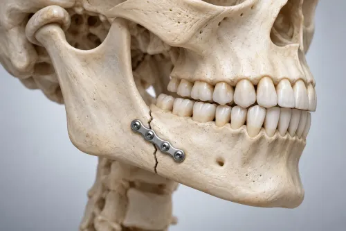
When the orbit, eyelid, cheek, jaw or frontal sinus is injured, ask how the teams will coordinate care. Emergency ophthalmology may need to assess the eye, an oculoplastic surgeon the eyelid, orbit or tear drainage system, and maxillofacial or ENT clinicians the associated facial or sinus injury.
The Face Clinic may be relevant when you need coordinated reassessment of these structures in Chennai; ask which clinician will examine the threatened structure and when reassessment occurs, not simply whether surgery is available.
Related services
OCCULOPLASTY AND FACIAL PLASTIC SURGERIES HEAD AND NECK TRAUMA MANAGEMENT ₹500 head and neck trauma associated with orbital fracture surgeries, facial fracture surgeries, secondary post traumatic deformity surgeries associated... View service → |
Maxillofacial Trauma & Reconstruction Facial trauma, whether from road traAic accidents, industrial injuries, sports, or assault, requires timely and skillful intervention to restore both... View service → |
Frequently asked questions
Which facial-trauma signs mean you should go to the emergency department?
Seek emergency assessment for reduced vision, a new relative afferent pupillary defect, a displaced eye, severe eye pain, worsening double vision or inability to move the eye normally.
Which eyelid cuts need an eyelid injury specialist?
Request specialist assessment for cuts crossing the eyelid margin, inner corner, tear-drainage opening, full eyelid thickness or the muscle that closes the eye.
When does an orbital fracture need surgery rather than observation?
Surgery becomes more likely when the fracture causes persistent double vision, restricted eye movement, a displaced or sunken eye, trapped tissue or significant orbital-volume change.
How should you monitor an injury after the first decision in Chennai?
Follow the documented review plan and return urgently for worsening vision, increasing pain, new double vision, vomiting, fever, a more sunken or displaced eye or difficulty moving it.
Related service
Our oculoplastic surgeons specialize in procedures involving the eye socket (orbit), eyelids, tear ducts, and adjacent face areas. View service → |
Keywords